

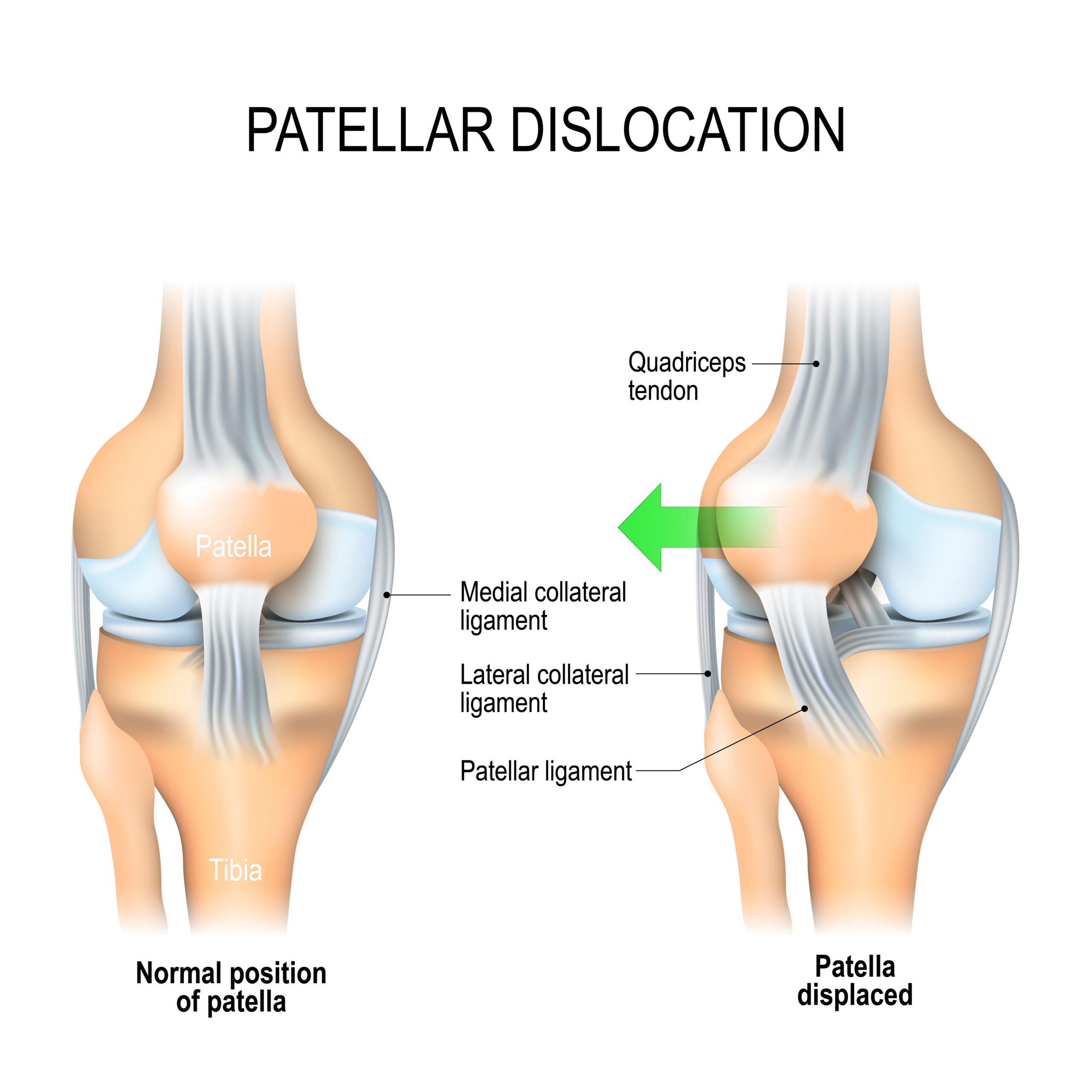
Patella instability occurs when the kneecap (patella) moves out of its normal position within the groove at the front of the knee. This may result in a complete dislocation, where the kneecap comes out of joint, or recurrent instability, where the kneecap repeatedly slips or feels as though it may give way.

Patella instability most commonly affects children, adolescents, and young adults, particularly those involved in sport. While some patients recover well following a first dislocation, others experience ongoing instability, pain, loss of confidence, and difficulty returning to sport.

Mr Jimmy Ng is a specialist sports knee surgeon in Nottingham with expertise in the assessment and treatment of patellofemoral instability, including MPFL reconstruction, tibial tubercle osteotomy, trochleoplasty, osteotomy (including derotation) and complex revision surgery.

What Is Patella Instability?

The patella normally glides within a groove on the femur called the trochlea. Several structures work together to keep the kneecap stable:

  • The shape of the trochlear groove
  • The medial patellofemoral complex including the medial patellofemoral ligament (MPFL)
  • The surrounding muscles, particularly the quadriceps
  • The alignment of the lower limb including rotational profile of the limb (twisting within the bone)

When one or more of these stabilising factors are abnormal, the kneecap can move excessively and may dislocate.

Patella instability exists on a spectrum ranging from occasional episodes of subluxation (partial dislocation) to recurrent complete dislocations.

Patella instability is often multifactorial.

Common risk factors include:

  • Trochlear Dysplasia: A shallow or abnormal trochlear groove provides less restraint to the patella.

  • Patella Alta: A high-riding kneecap engages later with the trochlear groove, increasing the risk of instability.

  • Increased TT-TG Distance: An increased tibial tubercle–trochlear groove (TT-TG) distance can create excessive lateral pull on the patella.

  • Generalised Ligamentous Laxity: Patients with hypermobility often have increased joint laxity and instability.

  • Lower Limb Malalignment: Femoral anteversion, external tibial torsion, valgus alignment, and rotational abnormalities can all contribute to recurrent instability.

  • Previous Patella Dislocation: A first-time dislocation frequently damages the MPFL, increasing the risk of future episodes.

Symptoms of Patella Instability

Common symptoms include:

  • Kneecap dislocation
  • Feeling that the kneecap is slipping or giving way
  • Pain at the front of the knee
  • Swelling after an injury
  • Clicking or catching sensations
  • Apprehension when changing direction or pivoting
  • Difficulty returning to sport

Many patients develop a fear of twisting or pivoting movements due to concerns about recurrent dislocation.

Assessment

A detailed assessment is essential to identify the underlying cause of instability and guide treatment.

During consultation, I will assess:

  • History of dislocation episodes
  • Sporting goals and activity level
  • Limb alignment
  • Patellar tracking
  • Joint laxity
  • Quadriceps strength
  • Evidence of cartilage injury

Investigations may include:

X-rays

To assess alignment and patella height.

MRI Scan

To identify:

  • MPFL injury
  • Cartilage damage
  • Osteochondral fractures
  • Trochlear dysplasia
  • Patella alta
  • TTTG

CT Scan

Occasionally required to assess rotational alignment and TT-TG distance in complex cases.

First-Time Patella Dislocation

Many patients can be successfully treated without surgery following a first-time dislocation.

Treatment may include:

  • Short period of bracing
  • Physiotherapy
  • Strengthening exercises
  • Activity modification

However, surgery may be recommended if there is:

  • Significant cartilage damage
  • Osteochondral fracture
  • High-risk anatomy
  • Recurrent instability after initial treatment

Recurrent Patella Instability

Patients who experience repeated dislocations are at risk of:

  • Progressive cartilage damage
  • Persistent pain
  • Loss of sporting function
  • Early arthritis

In these cases, surgical stabilisation may be recommended.

Surgical Treatment Options

Surgical treatment is tailored to each patient’s anatomy and risk factors. All risk factors or abnormal anatomy, if severe enough, needs to be addressed to achieve the best possible outcome in the long term.

MPFL Reconstruction (using quadriceps tendon)

The medial patellofemoral ligament is the primary soft tissue restraint preventing lateral patella dislocation.

MPFL reconstruction restores stability by reconstructing the damaged ligament using a tendon graft. I use a strip of the quadriceps tendon leaving it attached to the patella (kneecap) so no additional implants are required to secure the graft. The graft is then secured onto the femur (thigh bone) to provide stability. Occasionally a synthetic graft can be used in patients where quadriceps muscle activity / weakness is a concern.

This is the most commonly performed operation for recurrent patella instability, but it is rarely performed in isolation due to the other risk factors commonly involved.

Tibial Tubercle Transfer (TTT) aka Tibial Tubercle Osteotomy (TTO)

In patients with increased TT-TG distance or patella alta, repositioning the tibial tubercle can improve patellar tracking and symptoms such as instability and pain.

The tibial tubercle is detached from the tibia (shin bone) and repositioned based on pre-operative imaging (XR/MRI/CT). A small plate and screws are used to secure the tibial tubercle back onto the tibia to allow it to heal.

Use of a hinged knee brace is recommended for around 6 weeks when mobilising to protect the osteotomy but can be removed for sleeping, showering and doing exercises.

Groove Deepening Trochleoplasty (Bereiter’s)

For severe trochlear dysplasia, reshaping the trochlear groove may be considered.

This is typically reserved for selected patients with recurrent instability and significant anatomical abnormalities.

This is often combined with lengthening of the tight soft tissues causing dislocation and maltracking to ‘rebalance’ the patella and quadriceps tendon.

Osteotomy (including derotation)

Lower limb alignment such as valgus (knocked-knees) and rotational profile such as excessive femoral anteversion (inward facing of the thigh bone at the knee (distal femur) in relation to the hip (proximal femur) or tibial torsion (outward facing of the shin bone at the level of the ankle) can signficantly contributes to patella instability.

These abnormal alignments cause outward force on patella and abnormal movement when walking causing excessive pressure on the patella leading to pain and instability.

Cartilage Restoration Procedures

Cartilage injury frequently accompanies recurrent patella dislocation.

Large acute osteochondral fractures (cartilage injury) often require early surgery to re-attach the fractured fragment.

Cartilage restoration procedures may be required for chronic repeated injuries to the cartilage if appropriate along with stabilisation surgery.

Recovery After Surgery

Recovery depends upon the procedure(s) performed.

Typical recovery following TTT and MPFL reconstruction (most common combination) includes:

  • Immediate weight bearing
  • Early physiotherapy to control swelling, regain range of motion and strength
  • Brace use when mobilising only for 6 weeks
  • Running at approximately 3–4 months
  • Return to pivoting sports at 6–9 months

More complex procedures such as osteotomy may require longer rehabilitation.

Will I need surgery after my first patella dislocation?

Not necessarily. Many patients recover successfully with rehabilitation alone. Surgery is usually reserved for recurrent instability, significant cartilage injury, or high-risk anatomical factors.

Can I return to sport after MPFL reconstruction?

Most patients successfully return to sport following rehabilitation, although timing varies according to the individual and the level of sport.

Can recurrent patella dislocations damage the knee?

Yes. Repeated dislocations can cause cartilage injury and increase the risk of developing patellofemoral arthritis later in life.

Is physiotherapy important?

Absolutely. Physiotherapy plays a crucial role both before and after surgery to optimise strength, movement, and long-term outcomes.

Book an appointment

Contact the clinic today on 0115 937 7687 (Spire Nottingham Hospital)

Schedule a consultation to discuss your treatment options and find the best solution for your unstable kneecap.